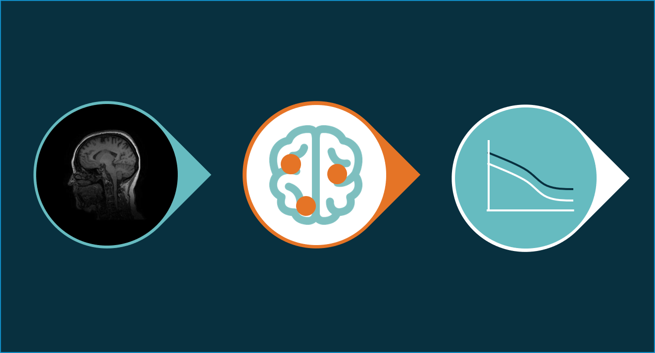
The field of radiomics covers a range of methods aimed at the extraction of quantitative features from medical images, and linking these features to pathological data. Developing algorithms to perform radiomics starts with collecting large amounts of medical images and clinical data. Subsequently, certain features (shapes, tissue texture, etc.) are extracted from the medical images, which thereafter can be linked with the clinical data, resulting in a radiomics training set. By presenting this combined training set to an artificial intelligence algorithm, the computer builds an understanding of the patterns that exist in how the medical images link to the clinical data, i.e. a radiomics model is developed.
Within the field of neuro-oncology, radiomics offers several opportunities to improve our understanding of brain tumors. As explained by Zhou et al. in a recent review paper, the deployment of radiomics to automatically analyze and identify gliomas, offers a wide range of possibilities. These include standardized tumor staging and more differentiated diagnosis, as well as refined monitoring of treatment response, e.g. following radiation treatment. Read the complete article:
https://www.quantib.com/blog/3-reasons-why-radiomics-can-revolutionize-brain-tumor-diagnosis-and-prognosis

